Functional images of the brain and psychopharmacology
Dr. Humberto Nicolini
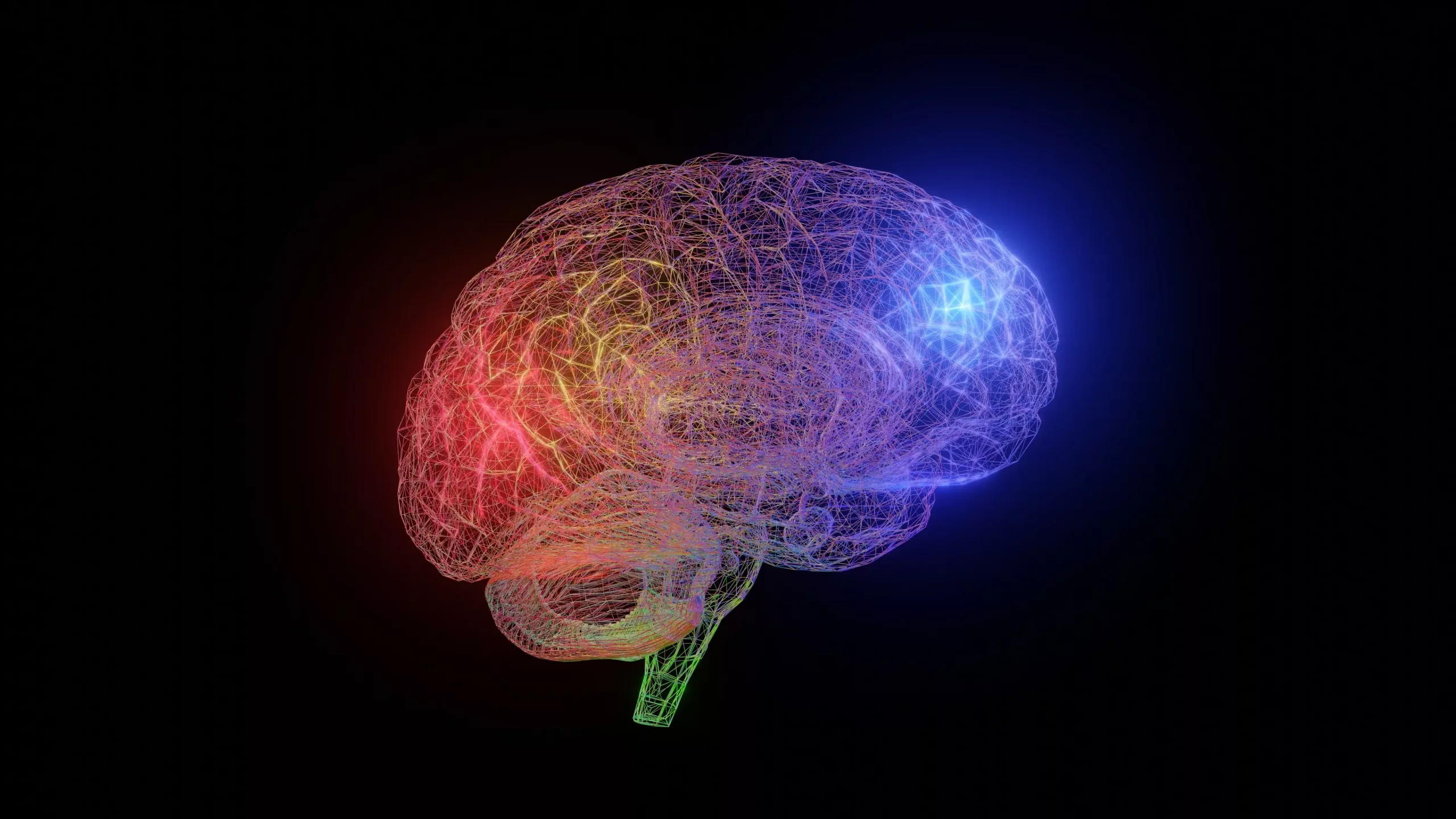
The various techniques of functional brain imaging have evolved surprisingly Ulti-mos in 15 years. Since the appearance of the skull radiography and electroencephalogram-techniques for over 100 years were the only tools for the brain, either in structure and function-until the advent of computers. This happens around the seventies, and when the revolution begins in the brain scan. Initially, the better appreciation of the brain through the use of computers in brain imaging is computed tomography (CT), which evolves into the reso-nance magnetic resonance (NMR) and finally the techniques and not simply but structural assessment of brain function. Within this category of imaging techniques fun-tions are the positron emission tomography (PET,
or PET in English), positron emission single photon (Tefu, or English SPECT) and functional magnetic resonance imaging (fMRI, or fMRI in English), which give beginning to the vision of the brain in action, both normal and pathological.
It is also necessary to mention the significant progress of neurophysiology through EEG
and novel computerized brain mapping techniques that will cause a later article. On the other hand, the parallel progress in psychopharmacology has been subject to the biochemical peripheral measurement techniques to quantify the effect of psi-codrugs metabolites. The major constraint was to see, in real time, how the brain interacted with Psicofarma-cos, and so not only inferential in peripheral or brain tissue. The appearance of PET and fMRI make this dream come true, and today it is possible to measure the molecular pharmacodynamics of psychotropic drugs, evaluating the occupation of the different specific receptors such drugs directly into the brain tissue of people receiving intake such substances.
Dynamic images of the brain have opened a new horizon “hypothesis testing” a pharmacological level as predicting drug response is one of the areas where the use of new technologies can ge-nerar immediate clinical knowledge. To illustrate this, we cite the classic work of Kapur and colleagues1, which, based on PET imaging, proposes the distinction between typical and atypical antipsychotics. This paper proposes that if an atypical antipsychotic, causing fewer extrapyramidal symptoms, because the receiver takes long enough to give an antipsychotic action and off of it before causing side effects (such as extrapyramidal symptoms). This theory was proposed based on the occupation of D2 dopamine receptors as measured by radioligand displacement of raclopride different antipsychotics, and where it was concluded that the bonding time and detachment from the receiver’s what antispsicóticos properly classifies as atypical. One-sequences with the rapid separation is resulting in that the receiver is again exposed to natural dopamine, before the next pulse of the drug. This bit of endogenous dopamine in the nigrostriatal system is sufficient for preventing the motor side effects caused by antipsychotics. However, as this example, south-giendo are many more in the field of psychopharmacology
for specific, such as social phobia, major depression, and obsessive-compulsive disorder, among others.2 In these conditions have been identified areas of the brain that predict response to antidepressants reuptake inhibitors of serotonin (such as the metabolic activity of the caudate nucleus). Moreover, recently it has also do Demostra-PET than placebo can activate the same brain circuits that analgésicos.3
Since the research that has generated the most important contri-butions in this field is Latin America, Dr. Nora Volkow, addictions deserve special atten-tion. By using both PET and fMRI, the work of Dr. Volkow have shown the involvement of dopamine reward systems and long-term damage in various addictions such as methane-fetaminas abuse, cocaine and binge eating.
Finally although these technologies are highly sophisticated and expensive in Latin America, we must not give us that-regardless of their understanding and practice, and they constitute one of the important pillars of theo-rich sustenance of the new psychopharmacology.
Bibliography
1. Kapur S, Seeman P. Does fast dissociation from the dopamine D2 re-
receiver Explain the action of atypical antipsychotics? A new hypothesis.
Am J Psychaitry. 2001; 158: 360-369.
2. Saxena S, Brody A, Ho M, et al. Differential cerebral metabolic Changes With paroxetine treatment of obsessive-compulsive disorder vs. major depression. Arch Gen Psychiatry. 2002; 59: 250-261.
3. C. Holden look alike Drugs and placebo in the brain. Science. 2002; 295: 947-948
4. Volkow N, Chang L, Wang G, et al. Association of dopamine transporter reduction With psychomotor impairment in methamphetamine abusers.
Am J Psychiatry. 2001; 158: 377-382.


Leave A Comment